Scoring tissue microarrays using Slide Score
Slide Score provides a fast way to score tissue microarrays using a familiar workflow with new possibilities for collaborations.
Scoring pathology slides with tissue microarrays (TMAs) can be a painful process. Pathologists using glass microscopes have a difficult time assessing the tissue cores and writing the scores down alone. Working with a resident or a PhD student who writes down the scores for them helps but introduces the need to book time to work together. Both methods risk skipping a row or a column and mismatching the sample identifier of the core.
Using digital pathology helps with scoring TMAs as pathologists have an easy overview of where in the slide they are and they can check the row and column of the core. However, proper support in software can improve it much further.
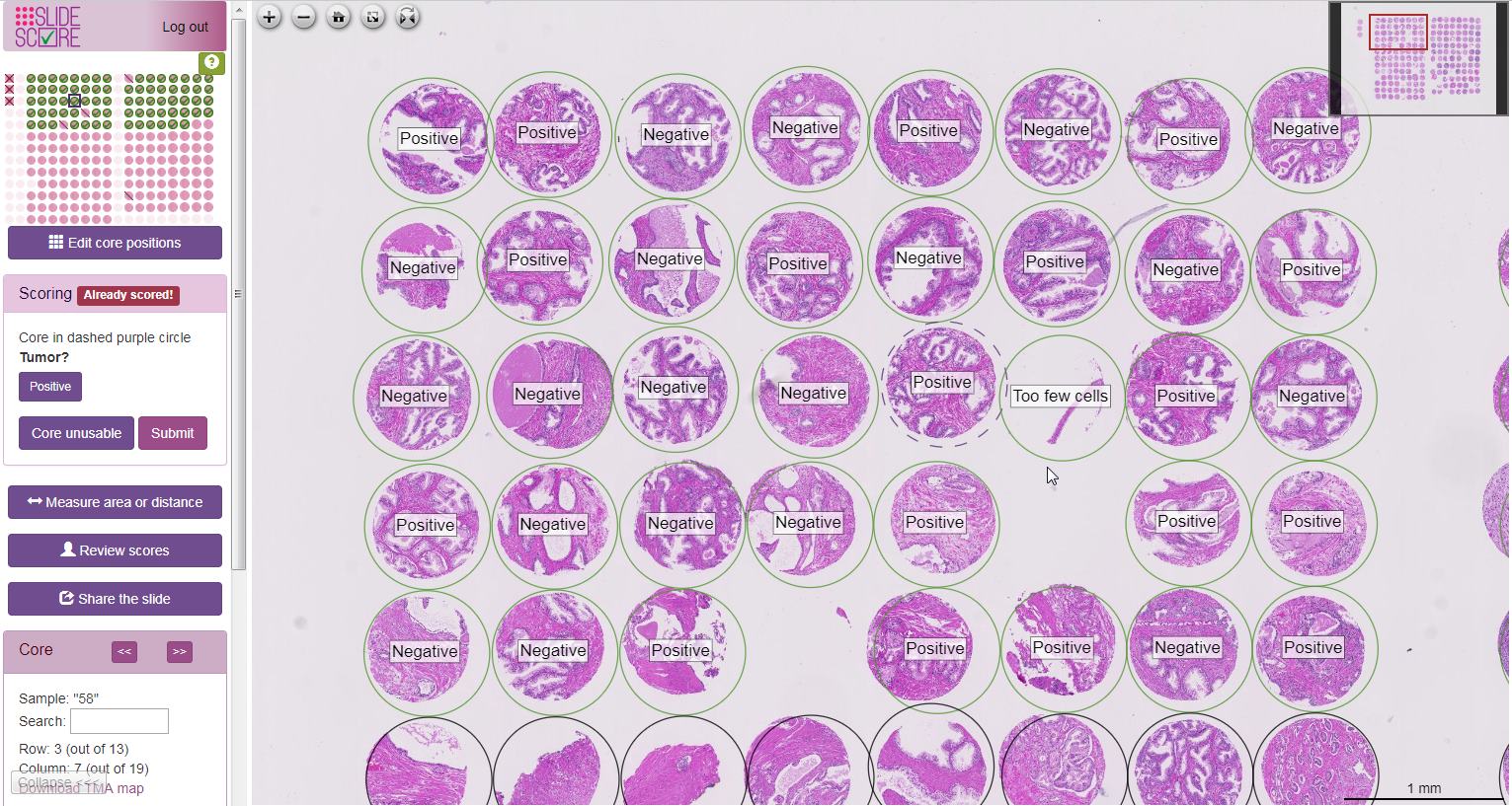
Slide Score supports reading tissue microarrays with a workflow that’s both more precise and faster than the previous methods. Immediately when opening a slide the viewer zooms to the first core that hasn’t been scored yet. If the scoring sheet has just a simple question (for example intensity, positive/negative or a percentage) pathologist just needs to click the button with the right answer and is instantly taken to the next core. At the same time a simplified TMA map (a grid with cores as they are on the slide including indication of control cores and holes) shows the progress with green checkmarks over completed cores.
Pathologists scoring TMAs with Slide Score regularly reach speeds of 1 TMA core/second for stainings where the assessment is easy. They can perform the scoring alone without having to schedule time with others and it’s simple enough to start that they can score some cores even if they have just 5 minutes of time between meetings.
To ensure that all cores are scored correctly pathologists can zoom out to view multiple cores and hold the Ctrl key to show an overlay with their answers. This makes it easy to spot any issues. It also allows for residents to perform the scoring first; resident pathologists can then easily retrieve and review their scores.
TMA blocks are often used for multiple stainings, if the serial sections align well it can be very helpful to view up the TMA cores at the same time. Synchronized viewing in Slide Score supports TMAs and allows viewing up to 8 slides with panning and zooming happening on all slides simultaneously.
It's possible to configure a Slide Score study to score all duplicates - TMA cores from a single sample - at once. If a block contains 3 cores from each sample the viewer will show all 3 at once and pathologist can choose the most representative, maximum or average answer depending on the methodology. This can save even more time!
If objective quantification is needed Slide Score integrates well with QuPath. Analysts can use a Slide Score plugin to open the whole set of TMA slides as a QuPath project and using a QuPath script analyze each TMA slide. They can retrieve the pixel coordinates of the TMA cores on the actual slide, run the image analysis workflow in QuPath and upload the results (for example annotation of tumor boundary, positive cells or just the percentage of positive cells in the TMA core) to Slide Score for a review by a pathologist.
Do you have a large set of TMA slides that needs to be scored? Slide Score offers affordable project-based pricing including hosting the slides!